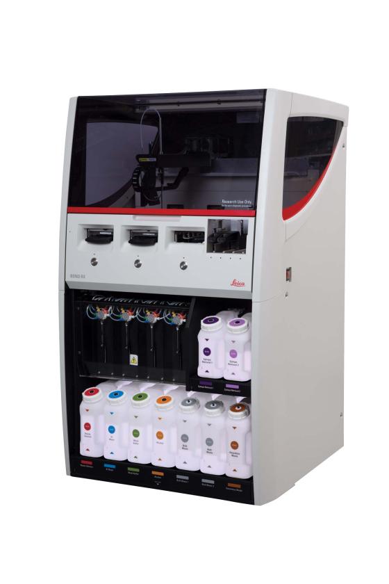
We offer immunohistochemistry (IHC) and immunofluorescence (IF) services employing the Leica Bond RX platform.
The process of immunohistochemistry involves the use of antibodies that are highly specific to target particular proteins of interest. These antibodies are labeled with a detectable marker, such as an enzyme or a fluorescent dye. When applied to tissue sections, these labeled antibodies bind to their target proteins, forming antigen-antibody complexes.
Subsequently, the tissues are treated with appropriate reagents that produce a visible signal, either through a color change (in enzyme-based techniques) or fluorescence (in fluorescent-based techniques) at the site of antigen-antibody binding. This signal allows researchers or pathologists to identify and localize the presence of specific proteins within the tissue sections under a microscope.
The QLMP can work up new antibodies to determine the appropriate conditions for staining. This involves staining 6 to 10 slides under different conditions to determine the optimal dilutions and antigen retrieval conditions.
Antibodies previously worked up at the QLMP:
- CD31
- Ki67
- CRLF2
- CD68
- ICAM1
- VE-Cadherin
- Anti-PKR
- FES
- Anti-FES
- Anti-Amyloid
- BaseScope (SRSF2)
