A healthy image
February 13, 2015
Share
Just as 3D technologies are revolutionizing the worlds of entertainment and printing, the power of 3D imaging is transforming health care.
For Amer Johri, assistant professor of echocardiography at Queen’s University and a clinician scientist at Kingston General Hospital, the rapid growth of 3D ultrasound imaging of the heart and vascular system opens up promising opportunities for advancing both patient care and doctors’ clinical skills.
Dr. Johri has made progress on both fronts. Returning to Queen’s in 2010 after completing an advanced fellowship in echocardiography at Harvard University Medical School, he created the Cardiovascular Imaging Network at Queen’s (CINQ) as a way to build existing, but disparate, pockets of strength in heart research into an investigative hub focused on imaging.
“I saw it as a home for people interested in cardiovascular imaging, as a way to share resources and expertise,” says Dr. Johri, a member of the KGH Research Institute who also holds the distinction of Fellow of the American Society of Echocardiography for his contributions to the field of ultrasound. “I knew what resources were available, what would work, and I had good relationships with the cardiologists. It was fun to start something from scratch.”
One of the centre’s significant areas of research is in the use of 3D ultrasound imaging of the carotid arteries, the major blood vessels in the neck, to detect heart disease. “Quantifying the buildup of the fatty deposits called plaque in the neck vessels can be a predictor of blockages elsewhere,” he explains.
It’s a relatively new area of research in which his group has already made an impact, he says. “Our results indicate that complete carotid ultrasound may serve as a simple, inexpensive, and low-risk test to rule out significant atherosclerotic cardiovascular disease.”
CINQ is also looking at measuring heart function through changes in the heart muscle not visible to the naked eye, using an advanced imaging technology known as “strain” or “speckle-tracking.”
A third study, conducted in collaboration with researchers at the Robarts Research Institute in London, Ontario, will incorporate 3D ultrasound into examining the effects of carnitine, a naturally occurring compound found in the body as well as in some foods, on patients with metabolic syndrome, the multiple conditions associated with heart disease that include obesity, diabetes, high blood pressure and high cholesterol.
The study was awarded a Heart and Stroke Foundation of Canada grant and also received support from the Department of Medicine and the Southeastern Ontario Academic Medical Organization (SEAMO).
A fourth area of research for Dr. Johri's lab is the study of the use of point of care ultrasound and development of training methodologies.
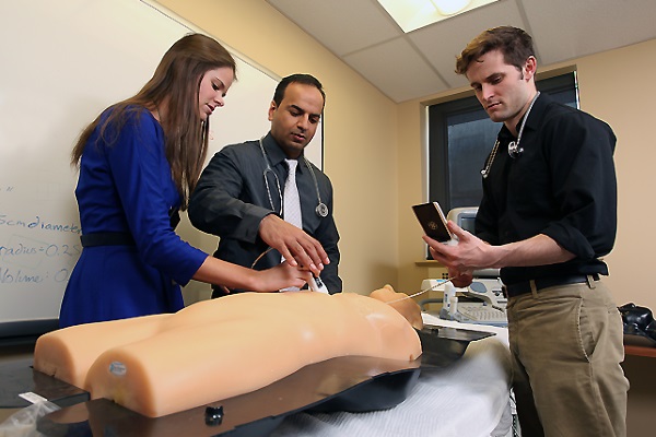
Advancements in ultrasound are also making a difference in how doctors examine their patients. In collaboration with Anthony Sanfilippo (associate dean, undergraduate medical education) the CINQ lab in 2010 began training medical students to use portable hand-held ultrasound during their physical exams of cardiac patients, making Queen’s School of Medicine one of the first to apply the emerging technology to clinical practice.
“All of the above are made possible because of KGH’s commitment to patient-oriented research,” says Dr. Johri. “It’s why I love the idea of the KGH Research Institute, because it supports the idea that research is important.”
You can follow Dr. Johri on Twitter @amerjohri.
This story is the fifth in a series on the KGH Research Institute, a collaboration between Queen’s and Kingston General Hospital, and the clinician-scientists recruited to work in the centre.